Overview
About This Condition
Retinal Vein Occlusion (RVO) occurs when one of the veins responsible for draining blood from the retina becomes blocked. When blood flow is disrupted, blood and fluid can leak into the retina, causing swelling and damage to the light-sensitive tissue at the back of the eye.
The condition can affect either the main retinal vein or one of its smaller branches. Depending on the location and severity of the blockage, vision loss may range from mild to severe.
RVO is one of the most common retinal vascular disorders and often occurs in people with underlying cardiovascular or vascular risk factors. Prompt evaluation and treatment can help preserve vision and reduce complications.
Retinal Circulation
When Blood Flow Is Interrupted, Vision Can Change Suddenly
Understanding symptoms and risk factors can help support timely care.
Symptoms
Blurred vision
Sudden vision loss
Dark spots or blind spots in vision
Floaters
Vision changes affecting one eye
Gradual or sudden decrease in visual clarity
Distorted vision caused by retinal swelling
Risk Factors
Diabetes
Glaucoma
High blood pressure
High cholesterol
Cardiovascular disease
Blood vessel disease
Conditions affecting circulation
Increasing age
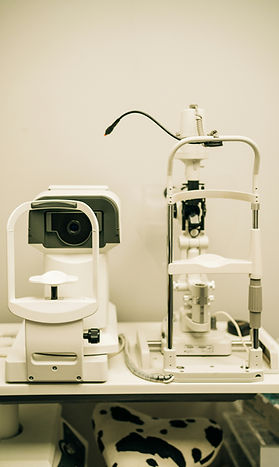
Diagnosis
How This Condition is Diagnosed
Retinal Vein Occlusion is diagnosed through a comprehensive retinal examination. Your ophthalmologist will examine the inside of the eye after dilating the pupils to evaluate the retina and identify signs of blockage, bleeding, or swelling.
Specialized imaging tests are commonly used to confirm the diagnosis and monitor treatment.
Optical Coherence Tomography (OCT) creates detailed cross-sectional images of the retina and helps measure retinal swelling.
Fluorescein Angiography uses a special dye and camera to evaluate blood flow within the retinal blood vessels and identify areas of blockage or leakage.
Treatment plans vary by patient and condition. Consult with your eye care specialist for personalized recommendations.
Treatment
Treatment Options
Treatment depends on the type of retinal vein occlusion, the severity of vision loss, and the amount of retinal swelling.
Treatment options may include:
Intravitreal medication injections to reduce retinal swelling
Anti-VEGF therapy to decrease fluid leakage
Laser treatment in selected cases
Monitoring for complications such as abnormal blood vessel growth
Management of underlying conditions such as diabetes and high blood pressure
The goal of treatment is to reduce swelling, preserve vision, and prevent further complications.